DICOM AI 3D VR viewer

Medical files massively used nowadays converted to 3D and furthermore enabled in virtual reality for in-depth examination.

This product virtualises DICOM scans in order to present 3D image to doctors. They can then examine the model of the human body in full 3D mode, not only on personal desktop but also via virtual reality glasses. Product enables in-depth exploration of organ condition and past changes examination along with patients.

Project

The initial need for the product appeared from a private medical institution in order to provide their patients with a truly visual and simply understandable way of their health condition. Specifically - DICOM scans. These scans are used widely in medical institutions around the world in order to present doctors the inner body scan of the patient - a precise organ.

- Easy and fast DICOM scan 3D viewer
- High fidelity 3D models of scanned organs with custom tools for in-depth examination
- Access patient DICOM scans directly from personal PACS
- Constant possible custom-made 3D environments and DICOM scans

Development time: 1 month
Engine: Unreal (UE4)
Platform: PC + HTC Vive
Visual style: realism
Language: EN (multi-language option available)
Target audience: surgeons and medical institutions
Stage: ready product

01- Research

Bringing 3D VR innovations to medicine sector

Medical systems nowadays still use regular 2D formats for organs' and other parts of body inspection. The barrier between immersive visualisations for patients and in-depth examination of organ remains. To introduce innovation to medical sector amongst other well-known virtual reality use cases (phobia treatment, surgery simulations, etc.) we have taken the daily use case of 2D DICOM scans and made them not only 3D, but 3D VR available. Multiple scans of one or several studies can be simultaneously opened in the same or different windows for comparison purposes in our product. In virtual reality the in-depth examination emerges to new levels. User can not only track the change process of the organ, but also apply different real conditions across certain period and foresee the possible changes.

The software has the capability to open and display studies obtained from different imaging modalities:
Digital Radiography (CR, DX)
Mammography (MG)
Computed Tomography (CT)
Magnetic Resonance (MR)
Positron Emission Tomography PET-CT (PT)
Ultrasonography (US)
Digital Angiography (XA)
Gamma Camera, Nuclear Medicine (NM)
Secondary Pictures and Scanned Images (SC)
Structured Reports (SR)

Many types of DICOM images are supported:
Monochromatic (e.g. CR, CT, MR) and color (e.g. US, 3D reconstructions)
Static images (e.g. CR, MG, CT) and dynamic sequences (e.g. XA, US)
Uncompressed and compressed (RLE, JPEG Lossy, JPEG Lossless, JPEG 2000)

The MPR tool provided within DICOM AI VR Viewer can be used to reconstruct images in orthogonal planes (sagittal, coronal, axial or oblique, depending on the base image plane). This can help to create a new perception of the anatomy that was not possible to visualize using the base images alone.

03- Maintenance

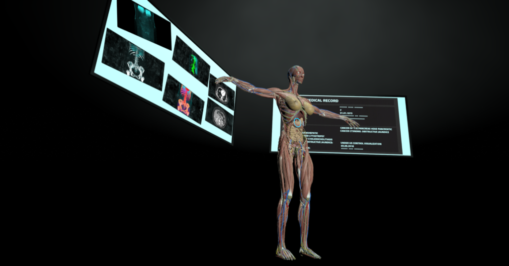
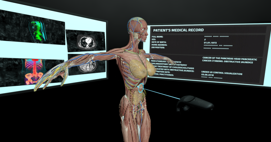
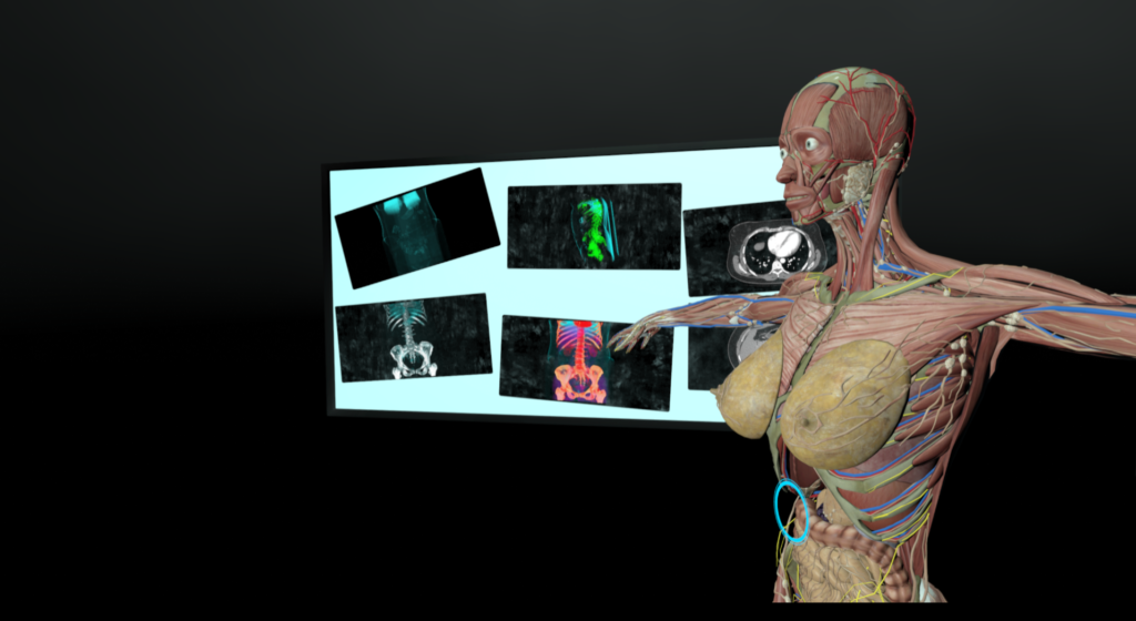
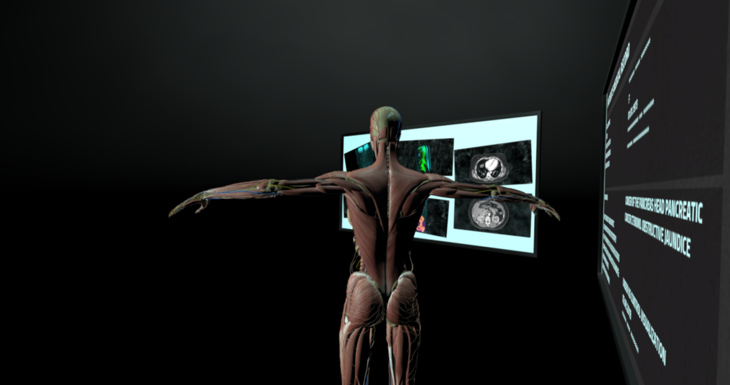
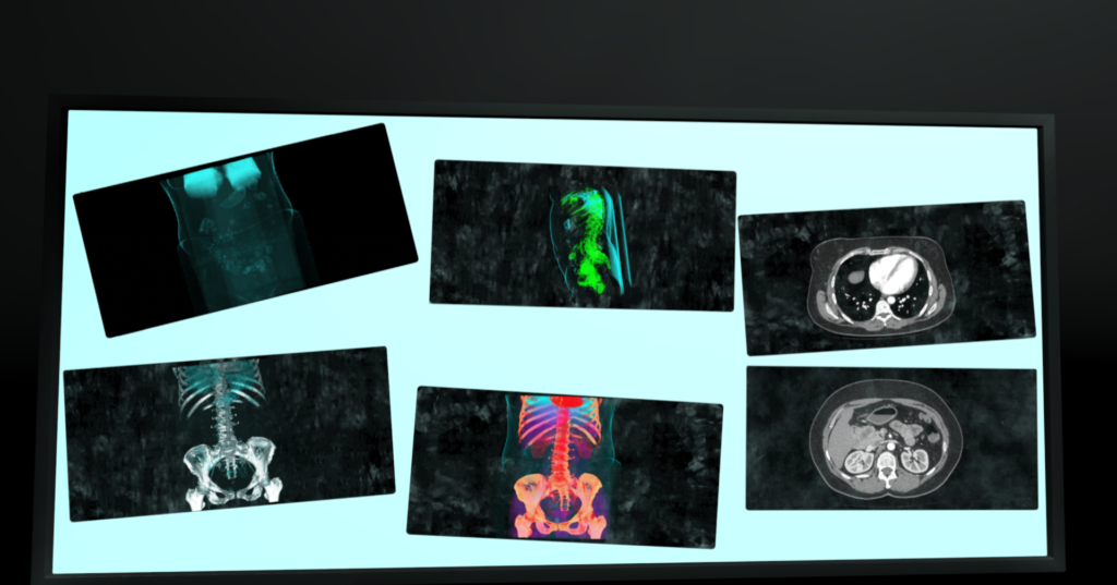
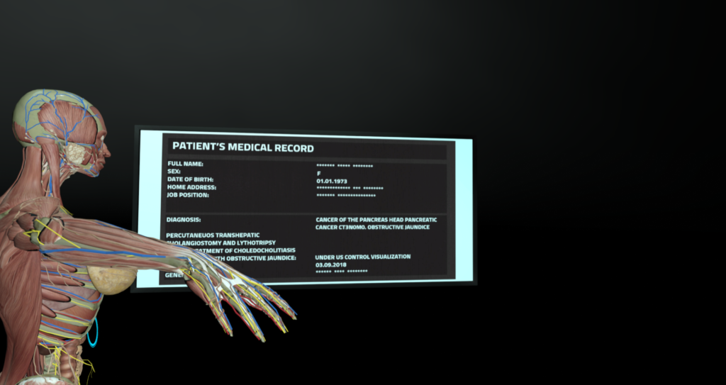
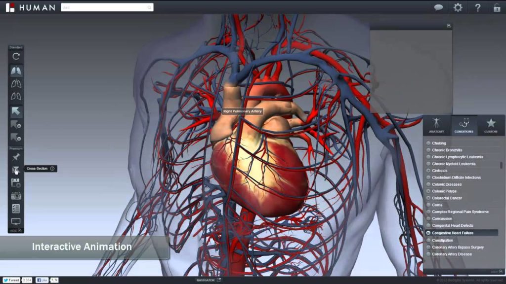
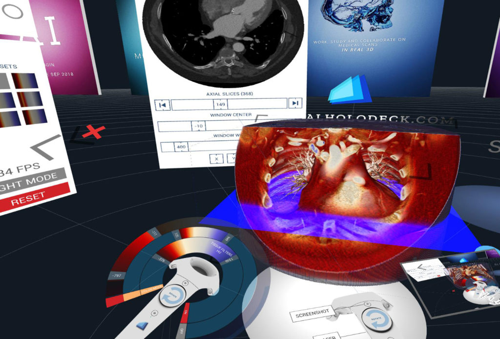
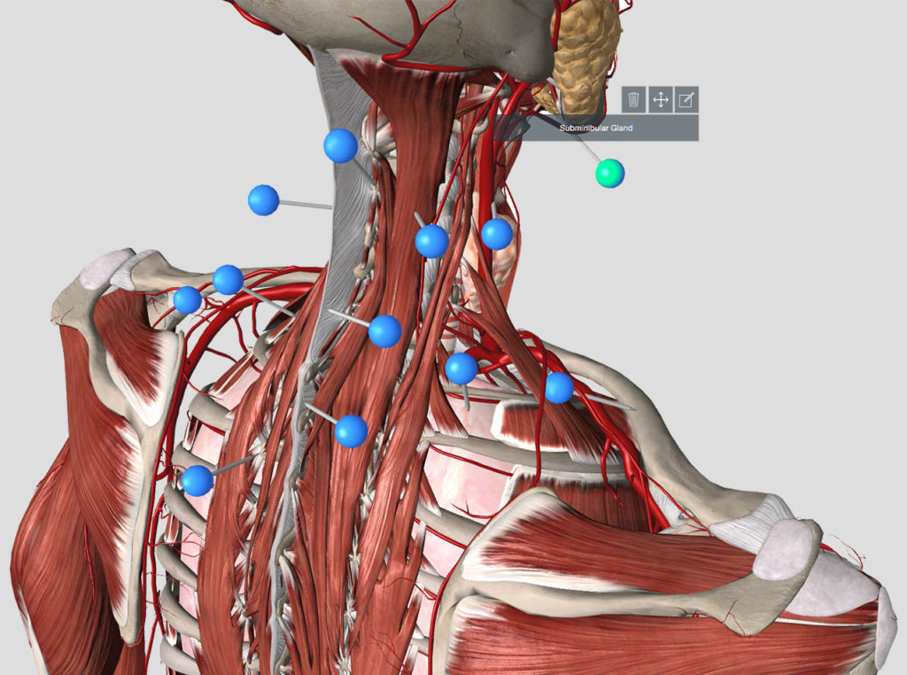
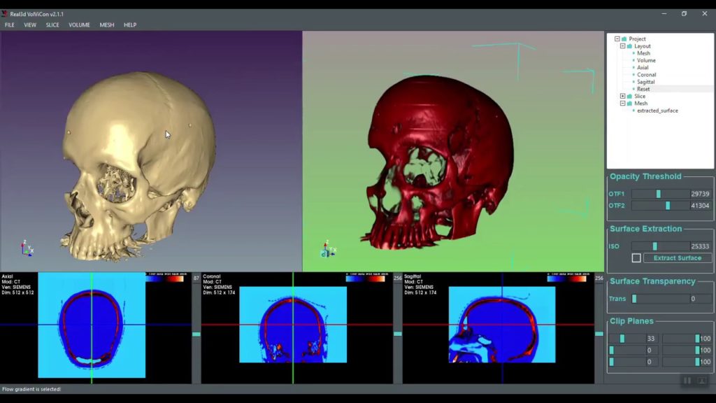
3D VR platform for all medical files

In our future updates for the product, we are planning to expand the database of scans while enabling other popular formats used in medical hardware nowadays. We are forming customisable 3D environments suited for custom-built doctor-to-patient virtual communication as well as implementing artificial intelligence (AI) based 3D model generation from 2D scans.

Any individual customisations are possible, so feel free to contact us upon your need.